Melanoma skin cancer can take many forms (in terms of size, shape, colour) and grow rapidly. Seeing melanoma images can assist you to identify the early stages of this type of skin cancer. In this blog post you’ll see different types of melanoma signs and symptoms, from early stage to advanced. You’ll also see what melanoma looks like on different parts of the body, from head to toe.
Do you want to read this article later?
Do you want to read this article later?
Thank you! Your submission has been received!
Oops! Something went wrong while submitting the form.
There are several different types of melanoma, including nodular melanoma, superficial spreading melanoma, lentigo maligna melanoma, acral lentiginous melanoma and amelanotic or ‘pink’ melanoma. Each form of skin cancer may develop differently. Looking at images of melanoma can provide a better understanding of the warning signs to look for during regular skin checks.
The most obvious melanoma warning signs are changes to existing moles or spots. They may alter in size, shape, colour, or in how they feel.
Usually, the most obvious melanoma warning signs are changes to existing moles or spots. They may alter in size, shape, colour, or in how they feel. For example, they may feel slightly raised. Melanoma can also appear as a new mole (more commonly in people aged 50 years or more).
Keep scrolling to melanoma skin cancer images for the five main types. However, remember that melanoma is only one form of skin cancer (albeit the most dangerous). Take a look at this handy guide to find out more about skin cancer.
Nodular melanoma images
Nodular melanoma is one of the most dangerous forms of melanoma. It accounts for about 15-20% of melanoma in Australia and New Zealand.
The most worrying thing about nodular melanoma is that it can grow fast: it is malignant from the time of appearance, which is why regular skin checks are so important.
As these melanoma pictures show, nodular melanoma is usually raised, often symmetrical, firm to the touch and grows or changes within a few months.
What does nodular melanoma look like?
Nodular melanoma is not necessarily dark or coloured. As the nodular melanoma images above show, the key features are that it’s raised, often symmetrical, firm to touch and, most importantly, changing or growing progressively.
In the early stages, you might not notice visible signs of change — perhaps the mole is itchy or just feels funny. This type of melanoma can affect anyone. Typically, it’s more common in men over the age of 50 and those with fair skin.
What's my skin cancer risk?
Answer six simple questions (takes less than 1 minute) to discover your risk and the right skin check for you.
Superficial spreading melanoma is the most common form of melanoma, accounting for around 70% of all cases. It tends to grow slowly and horizontally across the top layer of skin before moving to the deeper layers. It usually occurs on the back, chest and legs — areas that are all likely to get intense, periodic UV exposure from the sun. However, it can also appear in parts of the body that see little sun.
While it can affect people of all ages, this type of melanoma occurs most often in people in their 40s and 50s. Other risk factors include having fair skin, a lot of moles, a family or personal history of skin cancer, sunburn during childhood/adolescence, and having regular exposure to the sun or tanning beds.
As you can see in these melanoma skin cancer images, superficial spreading melanoma can be irregular in shape, variable in colour and similar to a freckle. It often appears on the legs, torso and upper back.
How do I detect superficial spreading melanoma?
Superficial spreading melanoma sometimes looks like a freckle, which can make it hard to identify, especially in the first stages. When checking your skin, look for these early signs:
Shape: Look for an irregular shape and borders. Superficial spreading melanoma can be raised or flat and can look like a freckle that is growing at its edges.
Colour: It may be brown, tan, black, red, blue and even white but usually has a combination of these colours.
Location: It usually appears on the torsos of men, the legs of women and the upper backs of both sexes — even in places that do not see the sun. It can appear in an existing mole or a new mole.
Changes: The mole or spot tends to change slowly, over the course of several years. It can sometimes feel itchy.
You can also use the ABCDEFG guidelines to help you identify the early signs of melanoma skin cancer. Early diagnosis is the key in successfully treating superficial spreading melanoma. So, if you notice any unusual spots on your skin, book a professional skin check straight away.
Amelanotic or ‘pink’ melanoma pictures
Just to make checking your skin even more difficult, there is a relatively uncommon type of superficial spreading melanoma that has no colour at all. Known as ‘amelanotic’ or ‘pink’ melanomas, these unusual spots have no melanin—that’s the dark pigment that gives most moles and melanomas their colour.
Amelanotic melanoma is no more dangerous than any other form of melanoma, yet its mortality rates tend to be higher than other types of melanoma. This is because it often goes undetected for longer, giving it more time to spread.
Image 1: an amelanotic, superficial spreading melanoma on the leg. Image 2: an example of a reddish-coloured amelanotic melanoma.
Amelanotic melanoma — what to look for
Amelanotic melanomas can be pink, red, purple, ‘normal’ skin colour, or even clear and colourless. They may even look just like a patch of abnormal skin. This makes them very easy to miss when self-checking your skin. However, there are other melanoma warning signs to look for. This includes a mole or spot that’s asymmetrical or has an irregular border. If a spot or mole appears suddenly or changes shape drastically, this may also be a sign of melanoma.
Sometimes, amelanotic melanomas resemble a tiny scar or acne that is healing. The biggest thing to look for is the ‘E’ for ‘evolution’ in the ABCDEFG guide — if you notice any changes in a mole or spot (no matter what the colour), seek a clinical diagnosis as soon as possible.
Lentigo maligna melanoma images
Lentigo maligna melanoma is the least common type of melanoma. It is a type of invasive skin cancer that accounts for around 5-15% of melanomas.
Lentigo maligna is a precancerous disease. It grows slowly and often stays on the outer surface of the skin. Although, if it starts growing into the second layer of the skin, it becomes the more malignant form, called ‘lentigo maligna melanoma’.
As this malignant melanoma image shows, lentigo maligna looks like a flat or slightly raised brown patch, similar to a freckle or sun spot.
What does lentigo maligna melanoma look like?
Both forms (precancerous and malignant) of lentigo maligna look like a flat or slightly raised brown patch, similar to a freckle or sun spot. They have a smooth surface and an irregular shape. While they are usually a shade of brown, they can also be pink, red or white.
They are usually larger than other types of skin cancer— often being at least six millimetres wide but can grow to several centimetres. This form of melanoma most often appears on the neck or face, especially on the nose and cheeks.
Keep an eye out for a mole with increased thickness, multiple colours (particularly black and blue), bleeding, itching or stinging. If you have any of these symptoms, get the mole or spot checked immediately.
Acral lentiginous melanoma pictures
Acral lentiginous melanoma (ALM) is a type of skin cancer that most commonly occurs on the palms of the hand or the soles of the feet. It can appear as a new spot or can develop within an existing mole.
As these melanoma skin cancer images show, acral lentiginous melanomas are usually brown or black, and occur on the palms of the hand, the soles of the feet and under fingernails or toenails.
What does acral lentiginous melanoma look like?
Often, acral lentiginous melanoma starts as a flat, slowly-growing patch of discoloured skin. Although it can also be reddish, orange or amelanotic in colour.
This type of melanoma is usually much darker than the surrounding skin (typically brown or black) and tends to have a sharp border between the dark skin and the lighter skin around it. This contrast in colour is one of the most noticeable symptoms of this type of melanoma.
Acral lentiginous melanoma is the most common type of melanoma in people with darker skin and those of Asian descent. Although all skin types can develop this form of skin cancer. It may be hard to detect in the first stages, when the patch of dark skin is small and looks like a stain or bruise. As with all types of melanoma, early diagnosis and treatment are essential to catch this rare form of melanoma before it spreads further.
It’s important to know the ABCDEFG rules when checking for early signs of melanoma.
Pictures of melanoma on different body parts
Now that you know what different types of melanomas look like, let’s look at pictures of melanoma skin cancer on different parts of the body.
Melanoma develops in different areas of the body in men vs women. In men, melanoma is most often detected on the back and chest, and in women, it’s most often found on the legs. But remember that melanoma can appear anywhere on the body, even where you least expect it — from your scalp to your torso to the soles of your feet (see 7 places you wouldn’t expect to find skin cancer).
These melanomas were all detected on different parts of the leg. In women, the legs are the most common site of melanoma.
A range of images of melanoma found on the face. Other types of skin cancer such as basal cell carcinomas and squamous cell carcinomas are also commonly found on the face.
These melanomas were all detected on different parts of the arm — arms tend to be exposed to the sun more than other parts of the body.
As these pictures show, melanoma is fairly common on the nose — a good reason to wear a hat when you’re out in the sun!
All of these melanomas were detected on the back. In men, melanoma is more likely to affect the chest and back.
What does skin cancer on the scalp look like?
The appearance of skin cancer on the scalp will actually vary greatly depending on the type of skin cancer it is.
Basal cell carcinoma (BCC) initially appears as a flat or raised spot that is usually pink in colour. These spots can bleed quite easily and may also be shiny, rough or crusty. They can also look like a slightly discoloured patch of skin. BCC is he most common form of skin cancer but it is also the slowest to spread. If you see something on your scalp that looks like one of the signs described above, book in for a skin check immediately.
Squamous cell carcinoma (SCC) is very common on the scalp. It appears as a rough or scaly patch on the scalp. It can also become raised, firm, red or even crusty over time.
Melanoma typically appears as a brown or black raised lump with dark, irregular colours and borders. But keep in mind that it can also look like a pinkish raised lump or mole that grows quickly in size. Scalp melanomas are very difficult to detect as they can be easily hidden by hair. This often leads to a delay in diagnosis, making them more lethal and aggressive than skin cancers elsewhere on the body.
Scalp melanomas are typically more lethal and aggressive than others, likely because they are easily hidden by hair and more difficult to detect and diagnose.
What does skin cancer on the lip look like?
SCC is the most common form of skin cancer to appear on your lips and will usually take the form of a scaly red patch that bleeds quite easily. Sometimes SCC can even appear as a painful ulcer or other non-healing sore. This form of skin cancer is more likely to appear on your bottom lip as opposed to the top.
BCC skin cancers are less likely to appear on your lips than other forms, but that doesn’t mean it’s impossible. If a BCC does appear, it’s typically more likely to do so on your upper lip. Pay attention to any pink spots that develop that are tender to the touch and bleed quite easily.
Melanomas can appear on your lip and are usually darker in colour. Never ignore a mole that changes in colour. It can help ensure early diagnosis.
Squamous cell carcinoma (SCC) is the most common skin cancer to appear on the lips, but basal cell carcinoma (BCC) and melanoma can also develop in this area too.
What does skin cancer on the feet look like?
The feet are typically the last place that people think to look when checking for skin cancer.
BCCs are the most common type of skin cancer to appear in this area and can look like a mole, a scar or even an unusual rough spot. These areas tend to bleed easily and are prone to becoming uncomfortable when you’re sweaty.
In the early stages, SCCs usually appear on the feet as a red or scaly plaque which is commonly referred to as Bowen’s Disease. This indicates that the skin cancer has not had the opportunity to develop into a full-blown SCC yet so there is still time to act.
Melanomas on the feet are generally rare, but this means that they are often sadly overlooked. Acral lentiginous melanoma is the most common form of melanoma to appear on your feet and usually involves the development of a light-coloured patch of skin. It can be very hard to see at first which is why having a trained professional check your feet for skin cancer is so vital.
When checking for skin cancer on the foot, be sure to check between the toes, under the soles and under the toenails.
Identifying the early signs of melanoma
Melanoma is the most serious form of skin cancer. It can spread and become life-threatening very quickly. But the good news is that if it’s found early, it’s almost always treatable and beatable. Knowing what a cancerous mole looks like and what to watch out for when checking your skin is essential.
Keep in mind that skin or mole changes aren’t often accompanied by pain, so early melanoma warning signs can go unnoticed. The best way to spot cancer early is to be vigilant about checking your skin regularly.
There are three simple guidelines to assist you when self-checking your skin. The ABCDEFG rule, the ‘ugly duckling’ method, and the Scan Your Skin rule. These provide a practical and handy guide when checking for early symptoms of melanoma.
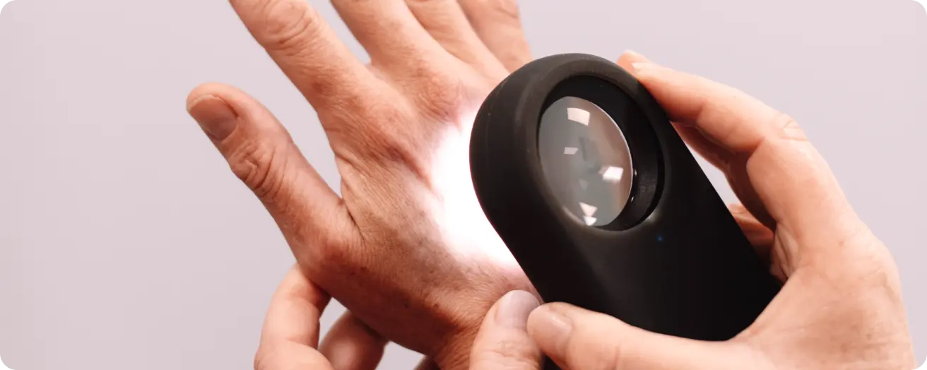
Regular skin checks are vital for monitoring any changes in your skin or early signs of skin cancer.
Melanoma can be difficult to spot in the early stages (especially to the untrained eye). A malignant melanoma can sometimes look very similar to a normal mole or spot.
MoleMap uses state-of-the-art imaging technology that can view both the exterior and dermoscopic (under the skin) structure of any mole. This, teamed with a dermatologist diagnosis, gives you the reassurance that any signs of melanoma or other skin cancers will be detected early.
MoleMap Team
At MoleMap we check, detect and treat skin cancer. Find out how you can protect your skin at your nearest MoleMap skin cancer clinic.
Oops! Something went wrong while submitting the form.
MoleMap are experts in skin cancer detection, diagnosis and proactive monitoring. Want the best protection against melanoma? Get your skin and moles checked early and often.