Superficial spreading melanoma accounts for around 70% of melanoma cases in Australia and New Zealand. Here’s how to spot this widespread skin cancer.
Typically starting out as a dark, pink or colourful spot with irregular edges, superficial spreading melanoma is the most common type of melanoma skin cancer. Find out the risk factors, what symptoms to watch out for and the survival rates.
Do you want to read this article later?
Do you want to read this article later?
Thank you! Your submission has been received!
Oops! Something went wrong while submitting the form.
Superficial spreading melanoma begins in the melanocyte cells. These are the pigment cells that live in the basal layer of epidermis (top layer of the skin). When the melanocytes grow uncontrollably, they can form cancerous tumours.
With Superficial spreading melanoma the malignant (cancerous) cells tend to remain in the epidermis for some time. When the cancerous cells remain at the site or origin, this is known as melanoma in situ. Superficial spreading melanoma grows outwards across the epidermis. It can remain in this phase for months to years.
How common is superficial spreading melanoma?
Superficial spreading melanoma makes up approximately 70% of all melanoma skin cancer cases. This type of melanoma is equally common in men and women. Superficial spreading melanoma can occur in adults of all ages. Yet only a small percentage (around 15%) arise under the age of 40. Even fewer (less than 1%) are found in people under the age of 20.
What's my skin cancer risk?
Answer six simple questions (takes less than 1 minute) to discover your risk and the right skin check for you.
Symptoms (See pictures of superficial spreading melanoma)
Superficial spreading melanomas can appear as a new lesion or grow within an existing mole. Symptoms of superficial spreading melanoma to watch for include:
A flat or slightly raised patch on the skin
An asymmetrical spot with uneven borders
A new or unusual lesion in different shades including light brown, dark brown, black, red/pink, blue or white
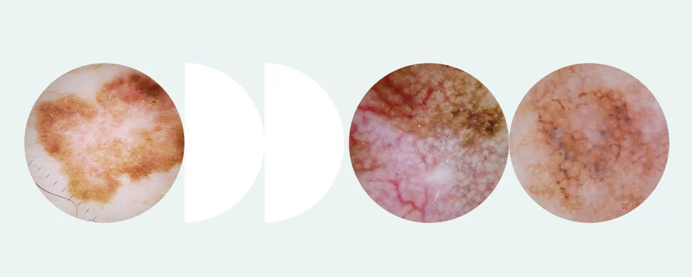
These melanomas can also be colourless—appearing a skin-tone spot. You may notice a mole or lesion with an unusual texture. Superficial spreading melanomas can incorporate a dark coloured nodular component. These superficial spreading melanoma images below may help you to spot this type of skin cancer. Learn more about how to detect melanoma symptoms.
Symptoms of superficial spreading melanoma to watch for include an asymmetrical spot with uneven borders.
Superficial spreading melanomas can appear as a new or unusual lesion in different shades including light brown, dark brown, black, red/pink, blue or white.
Superficial spreading melanoma can also present as a flat or slightly raised patch on the skin.
Who’s at risk?
A range of characteristics may increase your risk for superficial spreading melanoma. Key risk factors include:
Increasing age (it’s more common in people aged 40 years +)
UV exposure / history of sunburn
A large number of moles
Caucasian skin
Family history of melanoma
A previous history of skin cancer
How long does superficial spreading melanoma take to spread?
Unlike nodular melanoma, which quickly spreads, this type of melanoma grows slowly. Superficial spreading melanoma can take months, even years to invade other parts of the body. It spreads outwards across the skin before penetrating to the deeper layers.
How is superficial spreading melanoma diagnosed?
A biopsy is taken to diagnose superficial spreading melanoma. Typically, an excisional biopsy will be performed. This involves removing the entire lesion, including a margin of surrounding tissue.
A pathologist will examine the removed tissue under a microscope to determine if the cells are cancerous. The pathologist’s report will also include the Breslow thickness. This is a vertical measurement in millimetres from the top of the lesion to the deepest point of the tumour. It will show if the melanoma has spread (metastasised).
If it’s possible the melanoma has spread, a sentinel lymph node biopsy may be required. This involves removing some of the lymph nodes near the site for further investigation.
Typically, an excisional biopsy is taken to diagnose superficial spreading melanoma. This involves removing the entire lesion, including a margin of surrounding tissue.
Stages and treatment
Superficial spreading melanoma treatment will depend on the stage the skin cancer is at when diagnosed. Here’s an overview of the stages.
Stage 1
Nodular melanoma stage 1 refers to a thin melanoma (less than 2mm thick) that shows no sign of spreading (metastasis).
Stage 2
The nodular melanoma is more than 2mm in thickness. There is no evidence of spreading (metastasis).
Stage 3
Nodular melanoma has spread to nearby lymph nodes.
Stage 4
The nodular melanoma has spread to distant lymph nodes or skin.
Early stage superficial spreading melanoma (stages 1 and 2) is most often treated with surgery (biopsy/excision). The lesion is removed along with a 2 mm margin of normal tissue for diagnosis. This is always followed up by a second wider excision. The margin of this excision is determined by the depth of infiltration of the melanoma, as measured in the initial diagnostic excision.
For more advanced melanoma, where the cancer has spread, the following treatments may be used.
Immunotherapy uses medicines (immune checkpoint inhibitors)to assist the body’s immune system to find and kill the cancerous cells.
Targeted therapy used medication to block the growth of cancer by intercepting certain gene mutations in the melanoma cells.
Radiation therapy uses high-energy rays to destroy the cancer cells by damaging their DNA.
Chemotherapy is used for advanced stages of melanoma skin cancer. Anti-cancer (cytotoxic) medicines destroy the cancerous cells.
Superficial spreading melanoma survival rate
Compared with other types of melanoma, the survival rates for superficial spreading melanoma are high. A study of almost 100,000 superficial spreading melanoma cases found that the overall survival rate was 95%. Early diagnosis increases the chance of a positive outcome.
Prevention and early detection
The main way to prevent superficial spreading melanoma is to reduce your UV exposure. Following sun safety practices can help keep your skin healthy. This includes
Using sunscreen (SPF 50+)
Wearing a wide-brimmed hat
Covering your skin
Staying out of the sun when the UV index is at its highest (10am - 3pm)
In around 5-10% of melanoma patients the melanoma will recur. More than 20% of patients will develop a melanoma in situ (an unrelated melanoma). This is why regular skin checks are vital.
If you’ve already had melanoma, it’s likely your doctor will recommend how often you should get your skin checked. For people who haven’t had previous skin cancer, we recommend having a professional skin check at least yearly.
If you’ve recently noticed a new or suspicious spot and want to check it out quick, book a Skin Check. A melanographer (a nurse trained in skin cancer detection and triage) will examine your skin. Dermoscopic images will be sent for further review, and you’ll receive a full dermatologist report and diagnosis.
At MoleMap, our most comprehensive skin check is the Full Body MoleMap. It includes a thorough skin assessment with dermatologist diagnosis, total body photography, additional dermoscopic imaging of moles that may be at risk, and unlimited free spot checks for 12 months.
If you’ve recently noticed a new or suspicious spot and want to check it out quick, book a Skin Check. A melanographer (a nurse trained in skin cancer detection and triage) will examine your skin. Dermoscopic images will be sent for further review, and you’ll receive a full dermatologist report and diagnosis.
Oops! Something went wrong while submitting the form.
MoleMap are experts in skin cancer detection, diagnosis and proactive monitoring. Want the best protection against melanoma? Get your skin and moles checked early and often.